NRP 9th Edition · Condensed Reference
73-Card Study Guide
Every lesson of the NRP 9th Edition distilled into 73 reference cards. Tap any card to expand key facts, clinical tips and exam-relevant details.
Lessons 1–3: Foundations, Preparation & Initial Steps
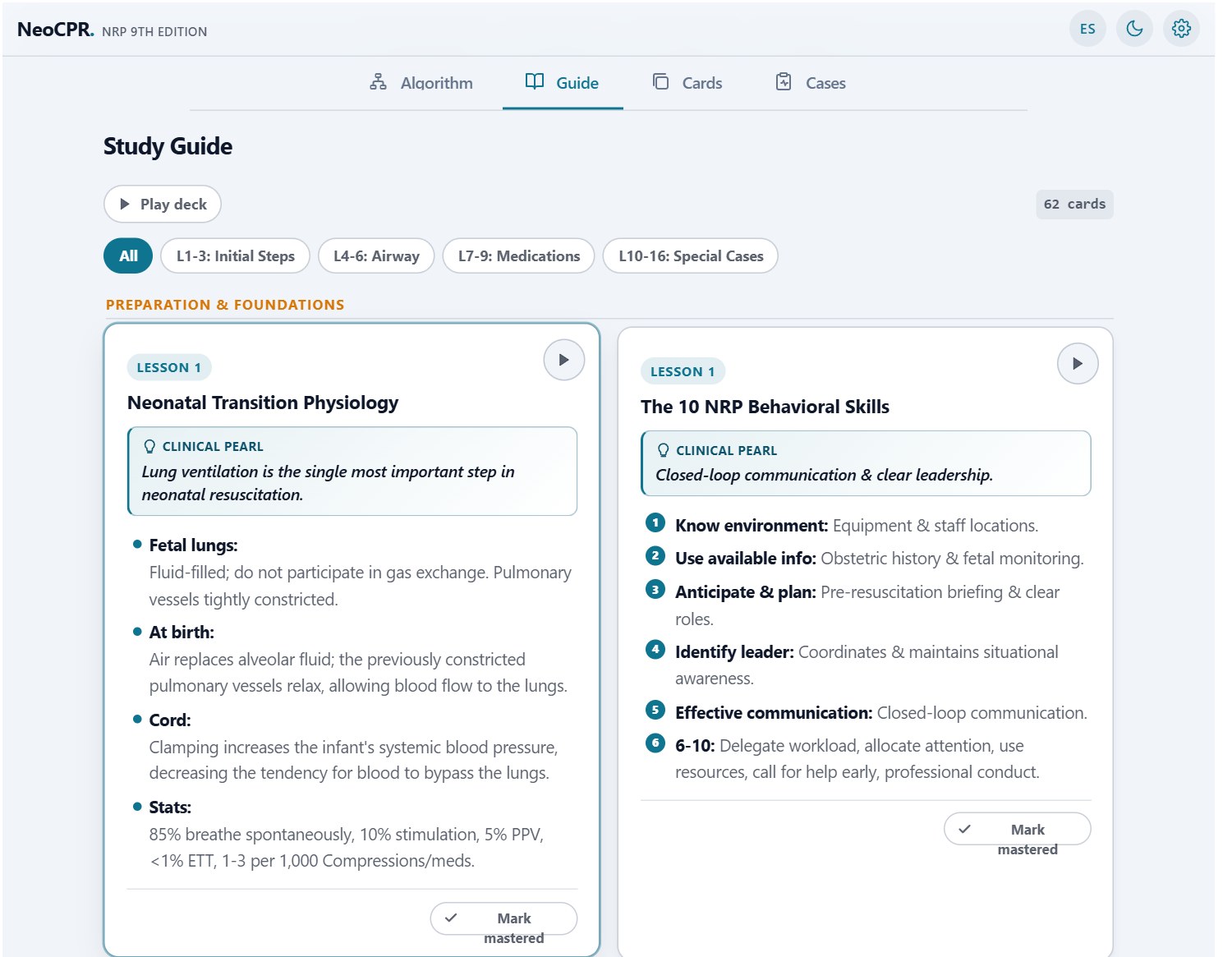
Lesson 1: Neonatal Transition Physiology
Lung ventilation is the single most important step in neonatal resuscitation.
- Fetal lungs: Fluid-filled; high Pulmonary Vascular Resistance (PVR).
- At birth: ENaC channels reabsorb fluid. Air stimulates eNOS → pulmonary vasodilation.
- Cord: Clamping increases SVR. SVR > PVR closes foramen ovale and ductus arteriosus.
- Stats: 85% breathe spontaneously, 10% stimulation, 5% PPV, <1% ETT, 1-3 per 1,000 Compressions/meds.
Lesson 1: The 10 NRP Behavioral Skills
Closed-loop communication & clear leadership.
- 1. Know environment: Equipment & staff locations.
- 2. Use available info: Obstetric history & fetal monitoring.
- 3. Anticipate & plan: Pre-resuscitation briefing & clear roles.
- 4. Identify leader: Coordinates & maintains situational awareness.
- 5. Effective communication: Closed-loop communication.
- 6. 6-10: Delegate workload, allocate attention, use resources, call for help early, professional conduct.
Lesson 2: The 4 Pre-birth Questions
Ask always before delivery occurs.
- 1. Expected gestational age? (Determines team & prematurity).
- 2. Is amniotic fluid clear? (Anticipate suctioning if meconium/obstruction).
- 3. Additional risk factors? (Preeclampsia, bleeding, general anesthesia, etc.).
- 4. Cord management plan? (Delayed clamping ≥ 60s vs immediate).
Lesson 2: Fetal Heart Rate Categories
Category III requires immediate intervention.
- Category I (Normal): FHR 110-160 bpm, moderate variability, no late/variable decelerations. Routine care.
- Category II (Indeterminate): Requires continuous monitoring and extra staff available.
- Category III (Abnormal): Absent variability with recurrent late/variable decelerations or bradycardia. Requires full resuscitation team at birth.
Lesson 2: 6-Area Equipment Checklist
Test suction at 80-100 mmHg and flowmeter at 10 L/min.
- 1. Warm: Radiant warmer, warm linens, hat, plastic wrap & thermal mattress (<32w).
- 2. Clear Airway: Bulb, 10F/12F catheter (80-100 mmHg), tracheal aspirator.
- 3. Auscultate: Neonatal stethoscope.
- 4. Ventilate: Blender, flowmeter 10 L/min, PPV, masks, 8F OG tube, LM size 1.
- 5. Oxygenate: Pulse oximeter, preductal sensor, SpO2 Table.
- 6. Intubate & Medicate: Laryngoscope (00,0,1), ETT (2.5-3.5), CO2 detector, Epinephrine 0.1 mg/mL, NS, UVC.
Lesson 3: Umbilical Cord Management
Avoid cord milking in <28 weeks due to severe intraventricular hemorrhage risk.
- Vigorous infants: Delay clamping for at least 60 seconds (increases iron & hemoglobin).
- Cord milking: Option for non-vigorous 35-42w. CONTRAINDICATED in <28w (causes IVH due to venous pressure surges).
- Immediate clamping: Placental abruption, bleeding placenta previa, cord avulsion, or non-intact circulation.
Lesson 3: 3 Initial Evaluation Questions at Birth
If all 3 are YES -> Routine care with mother.
- 1. Does the newborn appear term?
- 2. Good muscle tone? (Active flexion of extremities).
- 3. Breathing or crying? (Effective respiratory effort).
- 4. If YES to all 3: Skin-to-skin contact, warm, dry, position airway.
- 5. If NO to any: Move to radiant warmer for Initial Steps.
Lesson 3: Initial Steps under Radiant Warmer
Routine suctioning removed in 9th Edition. Do not suction without clear indication.
- 1. Warm: Radiant warmer (36.5-37.5 °C / plastic wrap <32w).
- 2. Dry: Remove wet linens to prevent evaporative cooling.
- 3. Position: Head in neutral sniffing position.
- 4. Stimulate if necessary: Gently rub back or soles.
- 5. Clear airway IF NEEDED: Wipe mouth then nose ONLY if secretions/obstruction present (NO routine suction).
Lesson 3: Preductal SpO2 Target Table
Sensor ALWAYS on right hand or wrist (preductal blood).
- 2 min: 65% - 70% | 3 min: 70% - 75%
- 4 min: 75% - 80% | 5 min: 80% - 85%
- 10 min: 85% - 95%
- 9th Ed Note: Minute 1 removed due to initial clinical imprecision.
Lesson 3: Free-Flow Oxygen & CPAP Indications
If HR >= 100 bpm but distress/cyanosis -> Try CPAP.
- Free-Flow Oxygen: Infant breathes spontaneously with HR ≥ 100 bpm, but SpO2 is below target. Titrate with blender at 10 L/min starting at FiO2 30%.
- CPAP: Infant breathes spontaneously with HR ≥ 100 bpm, but shows respiratory distress (grunting, retractions) or persistent hypoxemia. Start at 5-6 cm H2O.
Lesson 3: Meconium-Stained Amniotic Fluid (MSAF)
Routine tracheal suctioning is not indicated. The 9th Edition reaffirms immediate PPV initiation.
- Vigorous Newborn: Remain skin-to-skin with parent, dry, warm, and provide routine care regardless of meconium.
- Non-Vigorous Newborn: Move to radiant warmer for initial steps. Routine laryngoscopy/intubation for tracheal suction is NOT recommended.
- Tracheal Suction Indication: Only if airway obstruction is suspected and PPV fails to inflate the lungs.
Lesson 3: Hyperoxia Physiology & Oxidative Stress
Hyperoxia causes ROP, lung injury, and brain damage.
- Hyperoxia Risks: Production of reactive oxygen species (ROS) causing cellular damage, cerebral vasoconstriction, and retinopathy of prematurity (ROP).
- Physiological Titration: Start at FiO2 21% (≥35w), 21-30% (32-34w), or ≥30% (<32w). Titrate using a blender guided STRICTLY by the preductal SpO2 target table.
- Weaning: Gradually decrease FiO2 to room air once SpO2 stabilizes within the target range.
Lesson 2: Prenatal Risk Factors
More risk factors -> more staff present at birth.
- Maternal/obstetric: Preeclampsia/eclampsia, bleeding, emergency cesarean, forceps/vacuum, placental abruption, chorioamnionitis.
- Fetal/neonatal: Preterm (<36w) or post-term (≥41w), multiple gestation, macrosomia, meconium-stained fluid, FHR Category II/III, shoulder dystocia, cord prolapse.
- Medications: General anesthesia, magnesium sulfate, opioids within 4 hours before delivery.
Lesson 2: Resuscitation Team Staffing
The full team must be present AT BIRTH, not upon arrival.
- Every birth: At least 1 qualified person dedicated solely to the newborn.
- Risk factors present: At least 2 qualified people available.
- Anticipated complex resuscitation: Full team (≥4 people) with defined roles present at birth.
Lesson 3: Heart Rate Auscultation
6 seconds × 10 = beats per minute.
- Method: Auscultate the precordium on the left side of the chest.
- Calculation: Count beats for 6 seconds and multiply by 10 for bpm.
- Context: HR is the most important vital sign guiding resuscitation; assess it with breathing and tone.
Lessons 4–6: PPV, Intubation & Compressions
Lesson 4: Positive-Pressure Ventilation (PPV)
Success indicator #1: Rapid HR increase.
- PPV Indications: Apnea, gasping or HR < 100 bpm at 1 minute.
- Initial FiO2: ≥ 35w: 21%, 32-34w: 21-30%, <32w: ≥ 30%.
- Parameters: Flow 10 L/min, Rate 30-60 resp/min ("Breathe, two, three"). PIP 25 cm H2O, PEEP 5 cm H2O.
- OG Tube: Place 8F tube if PPV/CPAP continues for more than several minutes to decompress stomach.
Lesson 4: MR. SOPA Corrective Steps
Test PPV after M-R, S-O, and P.
- M: Mask adjustment | R: Reposition head.
- S: Suction airway (Mouth then nose) | O: Open mouth.
- P: Pressure increase (Increase PIP by 5 cm H2O up to max 40/30).
- A: Alternative airway (Insert ETT or Laryngeal Mask).
Lesson 5: Endotracheal Tube Selection Table
Laryngoscope in left hand, max time ~30 seconds.
- < 800 g (<26w): ETT 2.5 mm (2.0 mm optional) | Blade 00/0.
- 800 - 1,200 g (26-28w): ETT 2.5 mm | Blade 0/00.
- 1,201 - 2,200 g (29-34w): ETT 3.0 mm | Blade 0.
- > 2,200 g (>34w): ETT 3.5 mm | Blade 1.
Lesson 5: Tip-to-Gum Depth Table
Mark fixed strictly at upper maxillary gum line.
- 23-24w (<800g): 5.5 cm | 25-26w (700-800g): 6.0 cm
- 27-29w (900-1000g): 6.5 cm | 30-32w (1.1-1.4kg): 7.0 cm
- 33-34w (1.5-1.8kg): 7.5 cm | 35-37w (1.9-2.4kg): 8.0 cm
- 38-40w (2.5-3.1kg): 8.5 cm | 41-43w (3.2-4.2kg): 9.0 cm
- NTL+1 Formula: Nasotragus Distance (NTL) + 1 cm.
Lesson 5: Laryngeal Mask (Supraglottic Airway)
Excellent alternative if intubation or face mask fails.
- Indications: Inability to intubate or ventilate with face mask in infants ≥ 2,000 g (or ≥ 34w).
- Size: Size 1 (for newborns up to 5 kg).
- Technique: Guide with index finger along hard palate into hypopharynx until resistance is met. Inflate cuff with 2-5 mL air.
Lesson 6: Chest Compressions Technique & Ratio
Increase O2 to 100% and intubate before Compressions.
- Indication: HR < 60 bpm after 30s of effective PPV.
- Technique: 2 thumbs on lower third of sternum. Compressor at head of bed.
- Depth: 1/3 of anteroposterior chest diameter.
- Ratio: 3 compressions : 1 ventilation (120 events/min = 90 comp + 30 vent).
- Rhythm: "One-and-Two-and-Three-and-Breathe". Reevaluate HR after 60s with ECG.
Lesson 6: Pre-Epinephrine Compressions Checklist
Review before considering epinephrine.
- Compressions: 2-thumb technique on lower 1/3 sternum, depth 1/3 AP diameter.
- Airway: Endotracheal tube or Laryngeal Mask secured and verified with CO2 detector.
- 3:1 Ratio: 90 compressions + 30 ventilations = 120 events/min. Rhythm: One-and-Two-and-Three-and-Breathe.
- 100% FiO2: Maximum oxygen confirmed on blender.
- Vascular Access: Umbilical Venous Catheter (UVC) or Intraosseous (IO) line placed and ready.
Lesson 4: Colorimetric Exhaled CO2 Detector
Yellow = CO2 Present (Effective PPV/ETT).
- Yellow Color: CO2 successfully detected (effective alveolar ventilation or correct tracheal intubation).
- Purple / Blue Color: No CO2 detected (esophageal placement, obstructed tube, or zero cardiac output).
- False Positives: Epinephrine or surfactant contamination.
Lesson 4: Ventilation Devices Comparison
A self-inflating bag requires a dedicated PEEP valve to deliver PEEP or CPAP.
- T-Piece Resuscitator: Delivers consistent PIP & PEEP. Requires gas source. Enables CPAP.
- Flow-Inflating Bag: Delivers PEEP & CPAP. Requires gas source & mask seal.
- Self-Inflating Bag: Fills without gas. CANNOT deliver PEEP unless PEEP valve attached. CANNOT deliver CPAP or free-flow O2.
Lesson 4: 8F Orogastric Tube Measurement
Measure from nose bridge to earlobe to midpoint between xiphoid and umbilicus.
- Indication: PPV or CPAP lasting longer than several minutes (to decompress air-filled stomach).
- Measurement Technique: From bridge of nose -> Earlobe -> Midpoint between xiphoid process and umbilicus.
- Aspiration: Aspirate trapped air with 20 mL syringe and leave open to air.
Lesson 5: Laryngoscopy Anatomical Landmarks
Hold laryngoscope in left hand.
- Anatomical Sequence: Tongue → Vallecula → Epiglottis (above) → Glottis → Vocal cords in inverted "V" → Esophagus (below).
- Blade Tip: Place in vallecula (straight Miller blade) and lift along handle direction (DO NOT lever gums).
- Guided Depth: Insert ETT until vocal cord guide lines match cords.
Lesson 4: Post-PPV Reassessment (30s)
Rising HR is the best indicator of effective PPV.
- After 30s of effective PPV (chest movement): reassess HR and breathing.
- HR ≥100 bpm: PPV successful. Wean and adjust FiO2 per SpO2 table.
- HR 60-99 bpm: Continue PPV; reassess in 30s.
- HR <60 bpm: 30s more PPV via alternative airway (MR. SOPA); if still <60: 100% FiO2 and chest compressions.
Lesson 5: ETT Indications & Confirmation
Complete intubation in ~30s; beyond 30-50s may cause bradycardia.
- Indications: HR <100 not rising after optimized mask/LM ventilation; strongly recommended before compressions; tracheal suction (thick secretions); diaphragmatic hernia; surfactant administration.
- Primary confirmation: Exhaled CO2 turns yellow within 8-10 breaths + rapidly rising HR.
- Secondary confirmation: Symmetric axillary breath sounds; X-ray tip at T1-T2 (above carina).
Lesson 6: Post-Compressions Reassessment (60s)
Pause ~60s and assess HR (prefer cardiac monitor).
- After 60s of coordinated compressions with ventilation (3:1), pause briefly and assess HR.
- HR ≥60 bpm: Stop compressions; continue ventilation 30-60/min and adjust FiO2 per SpO2.
- HR <60 bpm: Continue compressions; give epinephrine IV/IO 0.02 mg/kg and secure emergency vascular access.
- PEA is treated as asystole. Rotate compressor after 2-5 min due to fatigue.
Lessons 7–9: Medications, Prematurity & Post-Resuscitation
Lesson 7: Epinephrine (IV/IO vs ET Dose)
Standard concentration: 0.1 mg/mL (1:10,000). Follow IV/IO doses with 3 mL normal saline flush.
- Indication: HR < 60 bpm after adequate PPV + 60s Compressions with 100% O2.
- IV/IO Route (Preferred): 0.02 mg/kg (0.2 mL/kg). Range 0.01-0.03 mg/kg. Rapid bolus + 3 mL NS flush. Repeat q3-5 min.
- ET Route (Provisional): 0.1 mg/kg (1.0 mL/kg). Range 0.05-0.1 mg/kg. Direct into tube WITHOUT flush, give PPV.
Lesson 7: Volume Expanders
Dose 10 mL/kg over 5 to 10 minutes.
- Indications: Hypovolemic shock (pallor, poor perfusion, weak pulses) or acute blood loss history.
- Solutions: 0.9% Normal Saline or Unmatched O-Negative Blood.
- Dose: 10 mL/kg IV/IO infused over 5 to 10 minutes.
Lesson 7: Emergency Umbilical Venous Catheter (UVC)
Insert only 3 to 4 cm until blood flash is seen.
- Technique: Cut cord 2 cm above skin. Identify single large vein (12 o clock position).
- Insertion: Advance 3.5F or 5F catheter only 3 to 4 cm (until blood return). Avoid deep liver advancement in emergency.
- Flush: Flush with 3 mL NS after epinephrine or expander.
Lesson 7: Inotropic & Vasoactive Medications (⚠️ Advanced NICU)
⚠️ Advanced NICU content. Not tested on the basic NRP exam.
- Dopamine: Continuous IV infusion 2 to 20 mcg/kg/min to improve cardiac output & BP.
- Dobutamine: Continuous IV infusion 2 to 20 mcg/kg/min if myocardial dysfunction predominates.
- Epinephrine Infusion: Continuous IV infusion (0.05 to 0.3 mcg/kg/min) in severe refractory shock.
- ⚠️ Note: These continuous infusions are advanced NICU management and are not part of the standard NRP algorithm.
Lesson 8: Preterm Resuscitation (<32w)
Polyethylene wrap without drying + Thermal mattress.
- Thermoregulation: Room temp 23-25 °C. Polyethylene wrap without drying + thermal mattress. Target axillary T° 36.5-37.5 °C.
- Breathing: Initial PEEP 5 cm H2O, PIP 20-25 cm H2O. Prioritize early CPAP (5-6 cm H2O) over prophylactic intubation.
- Initial FiO2: ≥ 30% in <32w.
- Neuroprotection: Avoid Trendelenburg, rapid volume boluses, and sharp PCO2 swings (prevents IVH).
Lesson 9: Therapeutic Hypothermia in HIE
Start within 6 hours of life in >=36 weeks.
- Criteria: Infant ≥ 36 weeks with perinatal asphyxia and moderate to severe HIE.
- Protocol: Cooling to 33.5 °C for 72 hours, initiated within 6 hours of life.
- Precautions: Avoid hyperthermia (worsens brain injury) and sodium bicarbonate infusions.
Lesson 9: Multi-System Post-Resuscitation Care
Unintended hyperthermia must be strictly avoided.
- Respiratory: Monitor pneumothorax, pulmonary HTN; consider surfactant; avoid unnecessary suction.
- Cardiovascular / Renal: BP control, urine output, perfusion. Consider inotropes if in shock.
- Metabolic: Glucose control (avoid hypo/hyperglycemia), electrolytes (Ca, K, Na).
- Bicarbonate: DO NOT infuse routinely (increases intracellular CO2 & IVH).
Lesson 9: Transport & S.T.A.B.L.E. Program
S.T.A.B.L.E. mnemonic for transport stabilization.
- S (Sugar): Blood sugar/glucose control.
- T (Temperature): Thermoregulation (avoid hyper/hypothermia).
- A (Airway): Secure airway & adequate oxygenation.
- B (Blood pressure): Blood pressure & tissue perfusion.
- L (Lab work): Basic laboratory evaluation.
- E (Emotional support): Family emotional support.
Lesson 9: Persistent Pulmonary Hypertension (PPHN)
Stability is the treatment: minimize handling.
- Definition: Persistently elevated pulmonary vascular resistance after birth; typically GA ≥34w.
- Presentation: Disproportionate hypoxemia with respiratory distress.
- Management: Avoid unnecessary suction, stimulation and bathing; minimize handling; O2 guided by preductal pulse oximetry; ventilatory support as needed.
- Monitor: Blood pressure and perfusion; rule out pneumothorax.
Lessons 10–16: Special Situations, Ethics & Quality
Lesson 11: Ethical Principles & Shared Decision Making
Parents serve as the primary surrogate decision makers.
- 4 Ethical Principles: Autonomy, Beneficence, Non-maleficence, and Justice.
- Parental Role: Primary surrogates for shared decision-making with medical team.
- Best Interest Standard: Decisions must always prioritize long-term newborn well-being.
- Ethics Committee: Consult if severe disagreements or irreconcilable uncertainty exist.
Lesson 11: 20-Minute Ethical Limit & Principle of Equivalence
20 minutes of 0 bpm = Ethical cessation of Compressions.
- 20-Minute Limit: If HR remains indetectable (0 bpm) after 20 minutes of effective Compressions, stopping resuscitation is ethically appropriate.
- Principle of Equivalence: Withholding initial resuscitation and withdrawing life support are ethically and legally equivalent.
- Trial of Therapy: If uncertain, start resuscitation, gather data, and transition to palliative care if non-responsive.
Lesson 11: Palliative & End-of-Life Care
Minimize suffering and provide compassionate family support.
- Humane Environment: Silence/remove noisy alarms and equipment. Gently clean baby and wrap in warm blanket.
- Family Contact: Encourage skin-to-skin holding and physical contact with parents.
- Pain Relief: Administer analgesia/opioids for comfort during agonal gasping.
- Declaring Death: Auscultate chest for at least 60 seconds to confirm absence of heart sounds.
Lesson 11: Prenatal Uncertainty, Organ Donation & Debriefing
Prenatal obstetrical margin of ±1 to 2 weeks.
- Obstetrical Uncertainty: 2nd/3rd trimester GA has ±1 to 2 weeks margin and weight estimates vary ±15-20%.
- Organ Donation: Contact regional procurement agency even if eligibility seems uncertain.
- Autopsy Option: Respectfully offer autopsy to clarify genetic/structural diagnoses.
- Team Debriefing: Conduct staff debriefing to process emotional impact & improve quality.
Lesson 10: Pneumothorax & Emergency Thoracentesis
Thoracentesis at 4th ICS anterior axillary line.
- Diagnosis: Sudden deterioration, asymmetrical breath sounds, positive transillumination.
- Puncture: 20G-24G catheter at 4th ICS (anterior axillary line) or 2nd ICS (midclavicular line), over upper rib margin.
- Aspiration: Connect 20-60 mL syringe with 3-way stopcock.
Lesson 10: Congenital Diaphragmatic Hernia (CDH)
Face mask positive pressure ventilation is contraindicated.
- Immediate Management: Immediate ETT intubation in delivery room. DO NOT use face mask.
- Decompression: Insert large OG tube (10F or Replogle) to continuous suction.
- Pressure Limit: Limit max PIP to 25 cm H2O to prevent hypoplastic lung barotrauma.
Lesson 10: Pierre Robin Sequence & Choanal Atresia
Pierre Robin -> Position in prone position.
- Pierre Robin: Position in prone position (brings tongue forward). Insert 2.5 mm ETT nasally into pharynx if needed.
- Choanal Atresia: Bilateral nasal obstruction. Insert ETT or McGovern nipple orally into posterior pharynx.
Lesson 10: Gastroschisis, Omphalocele & Myelomeningocele
Myelomeningocele: Use exclusively latex-free materials.
- Gastroschisis / Omphalocele: Enclose lower body in sterile plastic bag. Position on right side. 8F/10F OG tube to suction.
- Myelomeningocele: Position side or prone. Use protective donut ring. 100% LATEX-FREE materials.
- Opioid Depression: Naloxone CONTRAINDICATED. Provide PPV.
Lesson 12: Team Ergonomics & 5-Member Roles
Leader observes without getting trapped in manual tasks.
- Leader: Head of bed / oversees global scenario & delegates.
- Airway: Head of bed (PPV, intubation, UVC).
- Breathing Asst: Right side (SpO2, FiO2, timer).
- Circulation Asst: Left side (ECG, stethoscope, Compressions).
- Scribe / Recorder: Documents times, prepares drugs.
Lesson 13: Intraosseous (IO) Needle Outside Delivery Room
Insertion in anteromedial surface of tibia.
- Indication: Emergency vascular access outside delivery room if UVC unavailable.
- Insertion Site: Flat anteromedial surface of tibia, 2 cm below & medial to tibial tuberosity.
- SUPC: Sudden Unexpected Postnatal Collapse in first 12h during skin-to-skin or feeding.
Lesson 14: Quality Improvement (QI), SMART & PDCA
PDCA Cycle: Plan, Do, Check, Act.
- SMART Goals: Specific, Measurable, Achievable, Realistic, Timely.
- PDCA Cycle: Plan changes → Do/Implement → Check data → Act/Adjust.
- Balancing Measure: Ensures improvement doesn't cause unintended adverse consequences.
Lesson 15: Congenital Heart Disease & Prostaglandin E1
Target preductal SpO2 75%-85% at 10 min in CCHD.
- CCHD SpO2 Target: 75% - 85% at 10 min (avoid hyperoxia causing pulmonary steal).
- Prostaglandin E1 (PGE1): Continuous infusion at 0.005 to 0.03 mcg/kg/min to maintain ductal patency.
- HLHS / TGA with Restrictive Septum: Requires intubation, PGE1 & emergent balloon septostomy.
Lesson 16: NICU Resuscitation (NRP vs PALS)
NRP = 3:1 coordinated | PALS no ETT = 15:2.
- NRP in NICU: Indicated for respiratory cause. 3:1 ratio (90 comp + 30 vent/min coordinated).
- PALS in NICU: Indicated for cardiac/shock cause. No ETT = 15:2. With ETT = asynchronous (100-120 comp/min + 1 resp q2-3s).
- NICU Compressions: 1-2% of admissions receive compressions (10x more than delivery room).
Lesson 16: DOPE Airway Mnemonic
Review immediately upon sudden deterioration of intubated baby.
- D (Displaced): Displaced tube (check depth to gum & CO2 detector).
- O (Obstructed): Tube obstructed by mucus (suction).
- P (Pneumothorax): Pneumothorax or effusion (transillumination / POCUS).
- E (Equipment): Equipment failure or disconnection.
Lesson 16: Pericardiocentesis & Neonatal Arrhythmias
Unstable SVT -> Synchronized cardioversion 0.5 - 1 J/kg.
- Pericardiocentesis: 20G-24G subxiphoid catheter (0.5-1 cm below xiphoid) at 30-40° angle toward left shoulder.
- Unstable SVT: Synchronized cardioversion at 0.5 - 1 J/kg. (IV Adenosine 0.1-0.2 mg/kg if stable).
- VF / Pulseless VT: Defibrillation at 2 - 4 J/kg.
- Hyperkalemia: Primary metabolic cause of NICU arrhythmias/asystole.
Lesson 13: Home Birth & Transport
AAP/NRP do not recommend home birth (2-3× mortality).
- Home birth: AAP/NRP do not recommend it: 2-3× higher perinatal mortality.
- Resuscitation outside the delivery room: Same NRP principles; ventilation first.
- Self-inflating bag: 21% O2 without source; ~85-90% with O2 at 5-10 L/min. Laryngeal mask is the preferred alternative airway; tibial IO if UVC not feasible.
- Transport: Transfer any newborn requiring ventilation >30-60s.
Lesson 10: Pleural Effusion & Ascites
Suspect pleural effusion with respiratory distress + fetal hydrops.
- Pleural effusion: Suspect with respiratory distress and fetal hydrops. Drain at 5th-6th intercostal space, posterior axillary line.
- Ascites: Suspect with respiratory distress and abdominal distention. Paracentesis at right lower quadrant.
- General: Consider early intubation and coordinate with the neonatology team.
Algorithm Steps: The 18-Node Flowchart
Step 1: Equipment Preparation
4 pre-birth questions + 6-area checklist
- 4 Pre-Birth Questions: Expected age?, Clear fluid?, Cord plan?, Additional risk factors?
- 6-Area Checklist: Warmth, Airway, Auscultate, Ventilate, Oxygenate, Intubate/Medicate.
- Team Briefing: Assign roles (Leader, Airway, Compressions, Recorder) and test equipment before delivery.
Step 2: Delivery Moment
Mark delivery time (0:00) and evaluate vigor
- Minute Zero (0:00): Record exact time of complete expulsion and start resuscitation timer.
- Prevent Hypothermia: Receive in warm sterile towels or on mother's abdomen.
- Vigor Assessment: Rapidly observe respiratory effort, muscle tone, and initial vitality.
Step 3: Cord Management Plan
Delay clamping >=60s in vigorous infants
- Delayed Clamping (≥60 s): Indicated for vigorous and stable term and preterm infants.
- Immediate Clamping: Required if non-vigorous, or placental abruption/severe maternal bleeding.
- 9th Ed Golden Rule: Cord milking (UCM) is contraindicated in < 28 weeks due to severe IVH risk.
Step 4: Initial Evaluation (3 Questions)
Term? Tone? Breathing/crying?
- 1. Term gestation?: Determines lung maturity and need for radiant warmer.
- 2. Good muscle tone?: Active flexion of extremities and spontaneous movement.
- 3. Breathing or crying?: Effective respiratory effort without apnea.
- Rapid Triage: If YES to all 3 → Routine care with mother. If NO to any → Radiant warmer.
Step 5: Routine Care with Mother
Skin-to-skin, warm, dry, and observe
- Skin-to-Skin Contact: Place newborn on mother's chest or abdomen.
- Warm and Dry: Cover with warm blankets and hat; dry gently.
- Position Airway: Keep head in neutral sniffing position.
- Continuous Monitoring: Monitor breathing, color, tone, and temperature ongoingly.
Step 6: Initial Steps (Radiant Warmer)
Warm, dry, position, stimulate, clear airway if needed
- Warmth: Preheated radiant warmer (36.5-37.5 °C). Plastic wrap for <32 weeks.
- Dry & Position: Dry body, remove wet linens, position head in neutral sniffing.
- Tactile Stimulation: Gently rub back or soles to encourage breathing.
- Airway: Clear mouth then nose ONLY if secretions/obstruction present (NO routine suctioning).
Step 7: Heart Rate Evaluation
Auscultate for 6 seconds on left chest
- Preferred Method: Apical auscultation with stethoscope for 6 seconds (multiply beats × 10 = bpm).
- ECG Monitor: Indicated when auscultation is difficult or during PPV and Compressions.
- Critical Thresholds: HR ≥ 100 bpm (normal), HR < 100 bpm (requires PPV), HR < 60 bpm (Compressions/Epi).
Step 8: Respiratory Effort Evaluation
Detect apnea, gasping, or grunting
- Apnea / Gasping: Agonal or absent breathing is an immediate indication for PPV.
- Respiratory Distress: Expiratory grunting, intercostal retractions, or nasal flaring.
- SpO2 Monitoring: Place preductal pulse oximeter on right wrist/hand if cyanosis or distress.
Step 9: CPAP / Oxygen Application
CPAP 5-6 cm H2O if breathing with distress
- CPAP Indication: Infant breathes spontaneously with HR ≥ 100 bpm but has respiratory distress or low SpO2.
- CPAP Parameters: Initial pressure of 5 to 6 cm H2O via T-Piece or Flow-Inflating Bag.
- Free-Flow O2: Hold near nose at 10 L/min starting at FiO2 30%, titrating to target SpO2.
Step 10: Positive-Pressure Ventilation (PPV)
Start if apnea, gasping, or HR < 100 bpm
- Indications: Apnea, gasping, or HR < 100 bpm at 1 minute.
- Initial FiO2: ≥ 35w (21%), 32-34w (21-30%), < 32w (≥ 30%).
- Parameters: Rate 40-60 resp/min ("Breathe, two, three"). PIP 25 cm H2O, PEEP 5 cm H2O.
- Decompression: Insert 8F OG tube if PPV/CPAP exceeds 2-3 minutes.
Step 11: Post-Ventilation Evaluation (15-30s)
Check HR increase and chest movement
- 15-Second Check: If HR is increasing, PPV is effective. Continue for another 15s and re-evaluate.
- If HR NOT increasing but chest MOVES: Continue PPV for another 15s before next step.
- If HR NOT increasing & chest DOES NOT MOVE: PPV is ineffective. Immediately start MR. SOPA.
Step 12: Post-Resuscitation Care
Monitor glucose, T°, HIE & hypothermia
- Continuous Monitoring: Oximetry, ECG, and vital signs in NICU or intermediate care unit.
- Thermal & Glucose Control: Maintain normothermia (36.5-37.5 °C) and monitor blood glucose.
- Therapeutic Hypothermia: Assess for moderate/severe HIE in ≥ 36 weeks (cool to 33.5 °C within 6h for 72h).
Step 13: MR. SOPA Corrective Steps
Mask, Reposition, Suction, Open, Pressure, Alternative
- M & R: Mask adjustment | Reposition head (Neutral sniffing). Test PPV.
- S & O: Suction airway (Mouth then nose) | Open mouth. Test PPV.
- P: Pressure increase (Increase PIP by 5 cm H2O up to max 40/30 cm H2O). Test PPV.
- A: Alternative airway (Insert Endotracheal Tube or Laryngeal Mask).
Step 14: FC < 60 bpm Evaluation
If HR < 60 after effective PPV -> Compressions 3:1
- Effective PPV Confirmation: Confirm HR stays < 60 bpm DESPITE 30s of chest-moving PPV (preferably via ETT/LMA).
- Increase FiO2 to 100%: Increase oxygen concentration immediately to 100%.
- Vascular Access: Order immediate Umbilical Venous Catheter (UVC) or Intraosseous (IO) line.
Step 15: Chest Compressions (3:1)
90 comp + 30 vent/min (120 events/min) with 100% O2
- Preferred Technique: 2-thumb technique on lower third of sternum. Compressor positioned at head.
- Ratio & Rhythm: 3 compressions to 1 ventilation ("One-and-Two-and-Three-and-Breathe"). 120 events/min.
- Depth: 1/3 of anteroposterior chest diameter with full recoil.
- Coordinated Time: Perform uninterrupted Compressions for 60 full seconds before re-evaluating.
Step 16: Intubation / Laryngeal Mask
Secure advanced airway before Compressions
- Timing: Recommended before starting compressions or if mask PPV fails to expand lungs.
- Laryngoscope & ETT: Miller blade (00, 0, 1). ETT: 2.5 mm (<800g), 3.0 mm (1.2-2.2kg), 3.5 mm (>2.2kg).
- Laryngeal Mask: Effective alternative in ≥ 2,000 g / ≥ 34 weeks if intubation fails.
- Confirmation: Colorimetric CO2 detector (turns yellow) + bilateral axillary breath sounds.
Step 17: Post-Compressions Re-evaluation (60s)
Pause after 60s of coordinated Compressions for ECG
- ECG Monitor Check: Fastest and most accurate method to assess HR without prolonged Compressions pauses.
- If HR ≥ 60 bpm: Stop chest compressions and continue PPV at 40-60 resp/min.
- If HR stays < 60 bpm: Continue Compressions 3:1 with 100% O2 and administer IV/IO Epinephrine immediately.
Step 18: Epinephrine Administration
0.02 mg/kg IV/IO q3-5 min if HR stays < 60 bpm
- IV/IO Route (Preferred): Target dose 0.02 mg/kg (0.2 mL/kg of 0.1 mg/mL). Range 0.01 - 0.03 mg/kg. Rapid bolus + 3 mL NS flush.
- ET Route (Provisional): Dose 0.1 mg/kg (1.0 mL/kg). Range 0.05 - 0.1 mg/kg while securing UVC/IO access.
- Volume Expanders: 0.9% NS or O-Negative blood at 10 mL/kg over 5-10 min for acute blood loss or shock.
- Frequency: Repeat IV/IO epinephrine every 3 to 5 minutes if HR remains < 60 bpm.
Ready for more depth?
The full module adds 322 flashcards with spaced repetition, 10 clinical cases and a timed QBank.
This study guide covers the essentials. The complete NeoCPR module adds spaced repetition, scoring, a 45-question exam simulator and 7 additional clinical scenarios — for a one-time $29.
Get the full module — $29